An arrhythmia is a heart rhythm that is out of sync. People with arrhythmias have a faster or slower beating heart than those without the condition. Your heart may beat abnormally for a variety of reasons, and the treatment you receive depends on the underlying cause. If your heart is racing, you’re dizzy or lightheaded, or you’re experiencing chest pain, see your doctor right away.

There may be no signs or symptoms of arrhythmia if it is “silent”. You can tell if you have an irregular heartbeat during a doctor’s examination by checking your pulse or listening to your heart.

The following symptoms were experienced by the patients with arrhythmia.

• ++Palpitations++: A fluttering or “flip-flopping” heartbeat, or a feeling that your heart has skipped a beat.

• Your heart is thumping.

• Dizziness or a feeling of lightheadedness are among the most common symptoms.

• Breathlessness.

• Pain in the chest.

• Insufficiency or exhaustion (feeling very tired).

• Low ejection fraction or heart muscle weakening.


Arrhythmias are brought on by a variety of factors.

• Coronary artery disease can cause arrhythmias.

• The heart’s irritable tissue (due to genetic or acquired causes).

• Pressure in the arteries.

• Heart muscle changes (cardiomyopathy).

• Valve malfunctions.

• Blood imbalances in electrolytes, such as sodium or potassium.

• Heart attack-related injuries.

• The process of recovery following cardiac surgery.

• Other health issues.

In order to confirm the presence of an irregular heartbeat, some of the following tests may be performed:

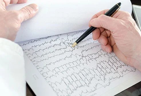
• Heart muscle electrical impulses are depicted on an electrocardiogram (ECG or EKG). Electrodes (small, sticky patches) are placed on the chest, arms, and legs to record an ECG on graph paper.

• Monitoring devices such as the Holter monitor.

• Arrhythmias that begin or worsen during exercise are recorded in a stress test, which is also known as an exercise stress test. An arrhythmia may be linked to underlying heart disease or coronary artery disease, which can be detected with this test.

• Any arrhythmia can be diagnosed with an echocardiogram, which is an ultrasound that shows the heart in order to check for any heart muscle or valve disease. With or without activity, this test can be administered.

• Percutaneous coronary angioplasty (PCA)

This procedure involves inserting a thin, flexible tube into the patient’s blood vessel and using an X-ray to guide it to the heart’s chambers. Through the catheter, a contrast dye is injected to produce X-ray images of your coronary arteries, heart chambers, and valves. This test can help your doctor determine if coronary artery disease is the cause of an arrhythmia. This test can also tell you how well your heart muscle and valves are functioning.

At Doral Health & Wellness Cardiology Center, we can help you find a solution for your heart problems, whether you believe they are caused by stress or are the result of underlying physical issues. As long as you’re concerned about your heart and want a second opinion, Doral Health & Wellness Cardiology Center is always ready and willing to help. Call us at 347-868-1012.

Leave a Reply

Your email address will not be published. Required fields are marked *

Arrhythmia

An irregular heartbeat is an arrhythmia (also called dysrhythmia). A regular heart rate is 50 to 100 beats per minute. Arrhythmias and abnormal heart rates don’t necessarily occur together. Arrhythmias can occur with a normal heart rate, or with heart rates that are slow (called bradyarrhythmias, less than 50 beats per minute). Arrhythmias can also occur with rapid heart rates (called tachyarrhythmias, faster than 100 beats per minute). In the United States, more than 850,000 people are hospitalized for an arrhythmia each year.

Many different factors may cause arrhythmias, including:
• Coronary artery disease.
• Electrolyte imbalances in your blood (such as sodium or potassium).
• Changes in your heart muscle.
• Injury from a heart attack.
• The healing process after heart surgery.
• Irregular heart rhythms can also occur in “normal, healthy” hearts.

irregular heartbeat - arrhythmia

The types of arrhythmias include:

Premature atrial contractions: These are early extra beats that originate in the atria (upper chambers of the heart). They are harmless and do not require treatment.

Premature ventricular contractions(PVCs): are among the most common arrhythmias and occur in people with and without heart disease. PVCs are the skipped heartbeat we all occasionally experience. It can be related to stress, too much caffeine or nicotine, or too much exercise in some people. But sometimes, PVCs can be caused by heart disease or electrolyte imbalance. A heart doctor should evaluate people who have a lot of PVCs and or symptoms associated with them. However, in most people, PVCs are usually harmless and rarely need treatment.

Atrial fibrillation:  is a prevalent irregular heart rhythm that causes the atria, the upper chambers of the heart, to contract abnormally.

Atrial flutter: is caused by one or more rapid circuits in the atrium. Atrial flutter is usually more organized and regular than atrial fibrillation. This arrhythmia occurs most often in people with heart disease and the first week after heart surgery. It often converts to atrial fibrillation.

Paroxysmal supraventricular tachycardia (PSVT): is a  rapid heart rate, usually with a regular rhythm, originating from above the ventricles. PSVT begins and ends suddenly. There are two main types: accessory path tachycardias and AV nodal reentrant tachycardias (see below).

Accessory pathway tachycardias: A rapid heart rate due to an extra abnormal pathway or connection between the atria and the ventricles. The impulses travel through the additional pathways as well as through the usual route. This allows the impulses to move around the heart quickly, causing the heart to beat unusually fast.

AV nodal reentrant tachycardia: A rapid heart rate due to more than one pathway through the AV node. It can cause heart palpitations, fainting, or heart failure. In many cases, it can be terminated using simple maneuvers, such as breathing in and bearing down, and others performed by a trained medical professional. Some drugs can also stop this heart rhythm.

Ventricular tachycardia (V-tach): A rapid heart rhythm originating from the heart’s lower chambers (or ventricles). The rapid rate prevents the heart from filling adequately with blood; therefore, less blood can pump through the body. This can be a severe arrhythmia, especially in people with heart disease, and may be associated with more symptoms. A heart doctor should evaluate this arrhythmia.

Ventricular fibrillation: An erratic, disorganized firing of impulses from the ventricles. The ventricles quiver and are unable to contract or pump blood to the body. This is a medical emergency that must be treated with cardiopulmonary resuscitation (CPR) and defibrillation as soon as possible.

Long QT syndrome: The QT interval is the area on the electrocardiogram representing the time it takes for the heart muscle to contract and then recover, or for the electrical impulse to fire impulses and then recharge. When the QT interval is longer than usual, it increases the risk for “torsade de pointes,” a life-threatening form of ventricular tachycardia. Long QT syndrome is an inherited condition that can cause sudden death in young people. It can be treated with antiarrhythmic drugs, pacemaker, electrical cardioversion, defibrillation, implanted cardioverter/defibrillator, or ablation therapy.

Bradyarrhythmia: These are slow heart rhythms, which may arise from the disease in the heart’s electrical conduction system. Examples include sinus node dysfunction and heart block.

Sinus node dysfunction: A slow heart rhythm due to an abnormal SA (sinus) node. Significant sinus node dysfunction that causes symptoms is treated with a pacemaker.

Heart block: A delay or complete block of the electrical impulse travels from the sinus node to the ventricles. The level of the block or delay may occur in the AV node or HIS-Purkinje system. The heart may beat irregularly and, often, more slowly. If severe, the heart block is treated with a pacemaker.

MEDICAL DISCLAIMER:

By reading this website, you acknowledge that you are responsible for your own health decisions. The information throughout this medical website is not intended to be taken as medical advice. The information provided is intended for general information regarding our cardiovascular clinic in Brooklyn, New York. If you experience chest pain, dizziness, nausea, or unusual shortness of breath, please stop and seek medical attention.

If you are interested in finding out more, please contact our Multi-Specialty Facility. Avoid worrisome self-diagnosis; the best cardiology doctors will properly diagnose your problem and refer you to a specialist if necessary. No information on this site should be used to diagnose, treat, prevent, or cure any disease or condition.